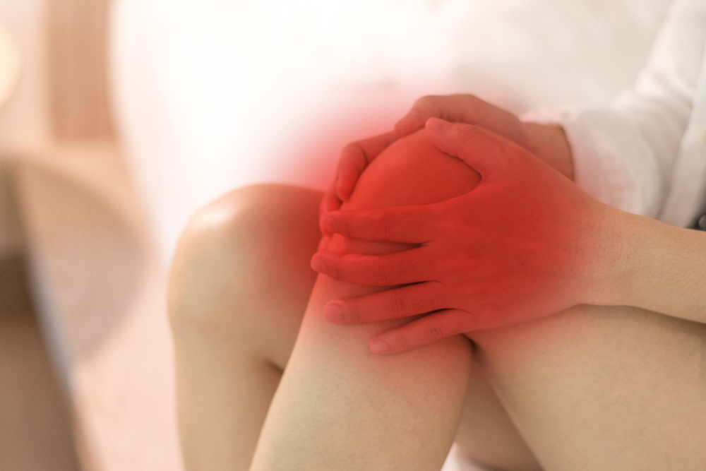
DIAGNOSIS
The attending doctor will check your medical history, particularly on your knee problems. The doctor will rotate and move the legs to check if there will be pain. Also, putting pressure on the knee like pressing and using a medical hammer will be helpful in diagnosing the syndrome.
To further determine the causes of pain in the knee, different tests are conducted. Here are as follows:
X-ray
This test helps visualize the bone structure. X-ray is an imaging technique that checks the bone but not the tissues around the bone.
CT Scan
The visualization of the bone and the soft tissues is possible through a CT Scan. The combination of the two imaging techniques delivers more efficient visuals on the bone and tissue condition of the knee.
MRI
Detailed images of the bones and the soft tissues are visualized through MRIs. The use of magnetic fields and radio waves enhances the view of the cartilages and ligaments of the knee.
MEDICATIONS
Most of the people suffering from patellofemoral syndrome try home treatments first to reduce mild pain. But, for severe knee pain, proper medical treatments are needed.
Home Treatment
Patellofemoral syndrome is commonly caused by inactivity. Due to this, proper and enough rest can help reduce the pain. Other simple yet effective treatments are as follows:
- The RICE method. This corresponds to the words rest, ice, compression, and elevation. Wrapping the knee with a bandage can help support the affected joint.
- A non-steroidal anti-inflammatory medicine can be taken to ease the pain.
- The use of orthotics or shoe inserts help stabilize the ankle and the foot which supports the knee.
- Massage can reduce the pain of the stressed muscles that causes the pain in the knee.
Medical Treatments
If the home treatments and medicines do not lead to pain reduction, consulting a doctor is needed. The usual recommendation is to have surgery. The surgical procedures that may be recommended to treat patellofemoral syndrome are as follows:
Arthroscopy. This is the insertion of a camera in the joint to see the damaged cartilage that must be taken out. It also refers to the release of tight tendons and the correction of the kneecap position.
Tibial Tubercle transfer. This process is the realignment of the kneecap. The tibial tubercle will be moved to the proper position.