LYMPHEDEMA

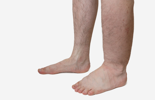
Swelling in one or more extremities that results from impaired flow of the lymphatic system.

  • Usually affects one arm or leg, but in uncommon situations both limbs are affected.
  • Primary lymphedema comes from a anatomical abnormality of the lymph vessels.
    • Rare and inherited.
    • Can occur on one or both sides of body.
  • Secondary lymphedema comes from an identifiable damage to or obstruction of normally-functioning lymph vessels and nodes.
  • Worldwide – Most commonly caused by filariasis (a parasite infection).
    • In the U.S., most commonly occurs in women who have had breast cancer surgery, particularly when followed by radiation treatment.
  • Usually occurs in one or both arms or legs, depending upon the extent and localization of damage.
    • May be only mildly apparent or debilitating and severe, as in the case of lymphatic filariasis (see above), in which an extremity may swell to several times its normal size.
    • First noticed as an asymmetry between both arms or legs or difficulty fitting into clothing or jewelry.
      • For severe swelling, fatigue due to added weight, embarrassment and restriction of daily activities may occur.
  • Non-pitting edema – firm, taut swelling that does not indent when pressed by a finger.
    • Due to long-term accumulation of fluid and proteins in the tissues that leads to inflammation and eventual scarring of tissues.
    • Peaud’ orange – thickening of skin with a lumpy appearance (orange peel effect).
    • The overlying skin can also become scaly and cracked, and secondary bacterial or fungal infections of the skin may develop. Affected areas may feel tender and sore, and loss of mobility or flexibility can occur.
  • Other symptoms:
  • Warmth, redness, or itching
  • Tingling or burning pains
  • Fever and chills
  • Decreased flexibility in the joints
  • Aching, pain, and fullness of the involved area
  • Skin rash
  • Suppresion of immune system
    • Leads to frequent infections and lymphangiosarcoma (malignant tumor of lymph vessels).

DIAGNOSIS

Physician may do/request:

  • History & Physical Exam
  • Imaging studies
    • CT
    • MRI
    • Lymposcintigrahy
    • Doppler Ultrasound


RECOMMENDED MEDICATIONS

  • No cure; treatment is for reduce swelling and control of discomfort
    • Compression treatments
      • Elastic sleeves or stockings
      • Bandages
      • Pneumatic compression devices
      • Manual compression
    • Exercise
    • Surgery for removal of excess fluids
  • Antibiotics for skin infections

Related Articles

TETRALOGY OF FALLOT

Overview and FactsTypes and SymptomsDiagnosis & MedicationsOverview and Facts Tetralogy of Fallot is a congenital heart defect that affects the [...]

TRICHINOSIS

Overview and FactsTypes and SymptomsDiagnosis & MedicationsOverview and Facts Trichinosis, also known as trichinellosis, is a parasitic infection caused by [...]

TRIGEMINAL NEURALGIA

Overview and FactsTypes and SymptomsDiagnosis & MedicationsOverview and Facts Trigeminal neuralgia is a neurological condition characterized by severe facial pain. [...]