COATS' DISEASE

COATS’ DISEASE

Coats’ disease is an uncommon condition wherein irregular veins in the retina expand and release liquid, bringing about harm to the retina and perhaps visual impairment. It frequently shows up at 8-10 years old, affects males multiple times more regularly than females, and routinely affects just one eye. The reason is obscure, and it doesn’t seem, by all accounts, to be innate.

The retina is the light-detecting layer of tissue in the rear of the eye. In Coats disease, tiny retinal arterioles and capillary tubes become spider veins that expand and shapes like little lights. These vessels release both liquid and fats, which develop in and below the retina. In previous cases, the liquid aggregation is sufficiently huge to disengage the retina. The retina’s broad sector may likewise lose appropriate blood supply, inciting the eye to create new vessels. These neovascular vessels can bring about additional issues, for example, glaucoma.

SYMPTOMS

Symptoms of Coats’ disease, for the most part, start in the early years. They might be moderate at the beginning, yet a few people may have serious indications immediately. Here are the early signs and symptoms:

  • Yellowish eye 
  • Strabismus eyes
  • Leukocoria, a white mass behind the focal point of the eye 
  • Loss of depth discernment 
  • Worsening of vision 

Later signs may include: 

  • Discoloration of the iris
  • Eye swelling
  • Retinal impartiality
  • Glaucoma
  • Deteriorate eyeball
  • Cataracts

These symptoms may commonly happen in one eye, even though both eyes are affected.

Coats disease stages

Coats’ disease is a persistent condition and is classified into five phases.

  • Stage 1

In the first stage, the ophthalmologist or doctor can notice unusual veins in your eye, but they have not begun to discharge.

  • Stage 2

The veins have started to discharge within the retina. If these discharges are minor, you can still have normal eyesight. With a broader issue, you might be encountering severe loss of sight.

  • Stage 3

Your retina is in moderation or fully disengaged.

  • Stage 4

You may experience severe eye pressure, known as glaucoma.

  • Stage 5

In earlier conditions, you may have entirely lost eyesight in the damaged eye. And also, you are at risk of having cataracts or atrophy of the eyeball.

DIAGNOSIS

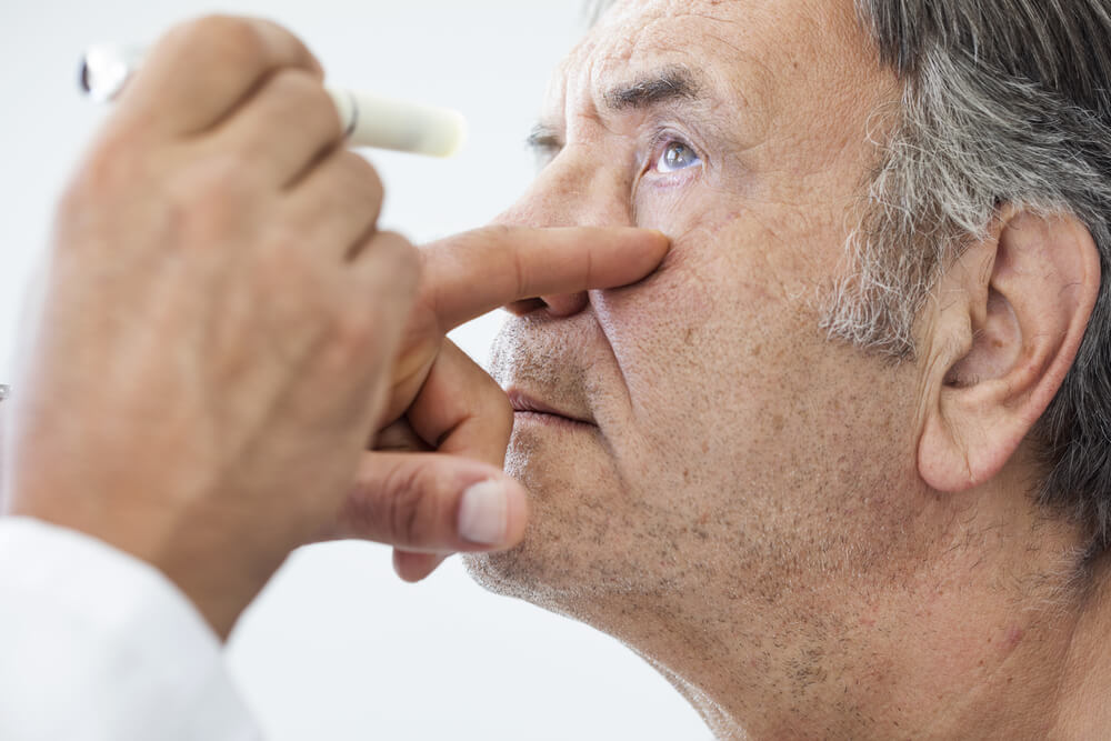
Specialists ordinarily diagnose Coats disease in young adults during a regular eye assessment after looking at the retina. Ophthalmologists utilize different imaging devices, including:

  • fluorescein angiography
  • optical cognizance tomography 
  • ultrasound to identify the degree of retinal implication 

TREATMENT

Sometimes, treatment of the disease may stop proceeding by itself and may get fixed. Still, some cases need treatment to prevent serious complications. A doctor may prescribe the use of freezing remedies or cryotherapy or laser to the discharge veins.

  • Steroids that manage the inflammation of the eye
  • Surgery to restore the parted retina

For severe cases, if the eye is already blind or painful, the doctor may require to remove it and change it with a prosthesis.

Related Articles

TETRALOGY OF FALLOT

Overview and FactsTypes and SymptomsDiagnosis & MedicationsOverview and Facts Tetralogy of Fallot is a congenital heart defect that affects the [...]

TRICHINOSIS

Overview and FactsTypes and SymptomsDiagnosis & MedicationsOverview and Facts Trichinosis, also known as trichinellosis, is a parasitic infection caused by [...]

TRIGEMINAL NEURALGIA

Overview and FactsTypes and SymptomsDiagnosis & MedicationsOverview and Facts Trigeminal neuralgia is a neurological condition characterized by severe facial pain. [...]