Seeing your GP
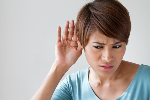
If ear problems such as hearing loss or persistent discharge occurs, consultation with your doctor is recommended.
The diagnosis of cholesteatoma maybe done after otoscopy. The procedure involves visualization of the ear with an otoscope, a handheld instrument that projects a beam of light.
If a cholesteatoma is seen, referral to a ear, nose and throat (ENT) specialist is done for further evaluation and management.
Treating a cholesteatoma
To confirm the diagnosis of cholesteatoma, the ENT specialist will re-examine the ear and may carry out some hearing tests. A computerized tomography scan (CT scan) is done to evaluate the extent of damage caused by the lesion.
Surgery
Surgery is done to remove the cholesteatoma, and is performed under general anesthesia. The surgeon makes an incision either behind or just in front and above your ear. As well as removing the dead skin cells, they may also need to remove some of the sponge-like mastoid bone (part of the skull behind your ear) and repair any hole in your eardrum.
After the surgery, the ear will be packed with a dressing which is left in place for a few weeks. Instruction regarding aftercare will be given.
The risks of surgery are similar to those of leaving the cholesteatoma untreated, such as hearing loss, tinnitus and vertigo, but generally the benefits of removing the cholesteatoma far outweigh the risks.
After surgery
Hospital admission is necessary at least overnight to monitor any immediate post-operative complications. The surgeon will advise you to have a week or two off work.
Self care advice
At home, make sure to keep the ear dry. Washing the hair may be done after a week provided that water does not get in the ear. This can be done by plugging the ear with a cotton wool with Vaseline. hen you get home, take care to keep the operated ear dry.
Swimming, strenuous activities and sports may need to be avoided for a few weeks. Check with the surgeon during follow-up visits regarding the resumption of these activities.
The surgeon may also advise avoiding flying for several weeks after surgery. Again, you can ask them about this at your follow-up appointment.
Follow-up appointments
If nonabsorbable sutures are used, the need to return for removal of stitches is usually after a week or two.
Most people have a follow-up appointment in a clinic within a few weeks of the operation, when any dressings in your ear will be removed.
A cholesteatoma recur, or may develop in the previously unaffected ear. Regular follow-up appointments are done to monitor any recurrence or growth.
Some people need a second operation after about a year to ensure that there are no fragments of cholesteatoma left behind.
When to seek medical advice
You should contact your GP or the ENT department of the hospital if you experience:
- discharge or significant bleeding from your ear or wound
- a high temperature (fever)
- severe or increasing pain
These problems could be a sign of a complication, such as an infection.