Introduction
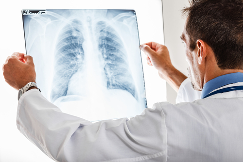
An X-ray is a quick and painless procedure commonly used to produce images of the inside of the body.It’s a very effective way of looking at the bones and can be used to help detect a range of conditions.
How X-rays work
- X-rays are a type of radiation that can pass through the body. They can’t be seen by the naked eye and you can’t feel them.
- Dense parts of your body that X-rays find it more difficult to pass through, such as bone, show up as clear white areas on the image. Softer parts that X-rays can pass through more easily, such as your heart and lungs, show up as darker areas.
When X-rays are used
Problems that may be detected during an X-ray include:
- bone fractures and breaks
- tooth problems, such as loose teeth and dental abscesses
- scoliosis (abnormal curvature of the spine)
- non-cancerous and cancerous bone tumours
- lung problems, such as pneumonia and lung cancer
- dysphagia (swallowing problems)heart problems, such as heart failure
- breast cancer
Preparing for an X-ray
- You don’t usually need to do anything special to prepare for an X-ray. You can eat and drink as normal beforehand and can continue taking your usual medications.
- For all X-rays, you should let the hospital know if you’re pregnant. X-rays aren’t usually recommended for pregnant women unless it’s an emergency.
Having an X-ray
- During an X-ray, you’ll usually be asked to lie on a table or stand against a flat surface so that the part of your body being examined can be positioned in the right place.
- The X-ray will last for a fraction of a second. You won’t feel anything while it’s carried out.
- While the X-ray is being taken, you’ll need to keep still so the image produced isn’t blurred. More than one X-ray may be taken from different angles to provide as much information as possible.
Contrast X-rays
In some cases, a substance called a contrast agent may be given before an X-ray is carried out. This can help show soft tissues more clearly on the X-ray.
- barium swallow– a substance called barium is swallowed to help highlight the upper digestive system
- barium enema– barium is passed into your bowel through your bottom
- angiography– iodine is injected into a blood vessel to highlight the heart and blood vessels
- intravenous urogram (IVU)– iodine is injected into a blood vessel to highlight the kidneys and bladder
Are X-rays safe?
- Generally, the amount of radiation you’re exposed to during an X-ray is the equivalent to between a few days and a few years of exposure to natural radiation from the environment.
- Being exposed to X-rays does carry a risk of causing cancer many years or decades later, but this risk is thought to be very small.
- The benefits and risks of having an X-ray will be weighed up before it’s recommended. Talk to your doctor or radiographer about the potential risks beforehand, if you have any concerns.


