RETINAL DETACHMENT

RETINAL DETACHMENT

The eye is filled with a gel (vitreous) that supports its shape. The retina is the light-susceptible tissue at the rear of your eye. It records visual images and sends them to your brain so you can see. Behind the retina is a thin layer of blood vessels that bring oxygen to the retina.

With age, the vitreous contracts, separating from the retinal tissue. When the vitreous separates, it causes “floaters” to appear. Floaters are small dots or strings that seem to be moving across your field of vision. Floaters are harmless. But they can be a sign that the retina has torn.

As the vitreous tears away from the retina, it may cause damage. If vitreous gel seeps through the crack and behind the retina, it can peel the retina away from part of its blood supply. The vision is restricted wherever the retina is disconnected. Retinal detachment can result in decreased vision, even if it is eventually repaired.

If the retina has torn but has not yet detached from the wall of the eye, treatment can create an adhesive scar around the retinal tear. This is like stapling around a hole in the wallpaper to keep it from coming off the wall. This can be done with laser treatment, which uses heat to create the scar. It can also be done with cryotherapy, which establishes an injury using cold. Eye surgery is needed to treat a retinal detachment. The type of surgery used depends on the type, size, and location of the detached part of the retina. Timely treatment is successful in over 90% of cases.

TYPES

There exist three kinds of  Retinal Detachment

Rhegmatogenous retinal detachment is the most common type of squad. It is caused by tears or holes in the retina called retinal breaks. A retinal break grants liquid from the center of the eye to experience the damage and settle under the retina. As the cash amasses under the retina, it drives the retina away from the layer underneath it.

Traction retinal detachment does not cause a specific tear or break in the retina. It takes place when scar tissue or other rare tissue is rooted on the outside of the retina, dragging the retina off from the layer underneath it. The leading cause of this condition is proliferative retinopathy, a condition most frequently caused by diabetes.

Exudative retinal separation evolves when blood or liquid from the center layer of tissue that forms the eyeball (choroid) streams into space under the retina and isolates the retina from the layer underneath it. The detachment does not involve tears in the retina or traction from the vitreous.

Symptoms of retinal detachment include:

  • Blurry eyesight
  • Bright flashes of light, especially in peripheral vision
  • New floaters in the eye that appear all of a sudden
  • Shadowing or decreased peripheral vision that seems like a curtain or shade across your vision
  • Sometimes there is no pain in or around the eye.

DIAGNOSIS

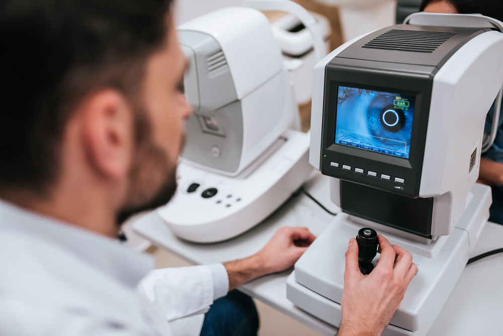
Ophthalmologists and optometrists will distinguish any retinal separations by looking at the rear of the eye with a slit lamp or an ophthalmoscope. The various kinds of retinal detachment are isolated dependent on the harm caused to the retina. A detached retina can be created, for instance, when disturbance begins in the vitreous gel, the clear part of the eye that fills your eye gets watery with age. Hence, it can enter between the retina and the layer underneath, making your retina detach. This kind of retinal detachment is called posterior vitreous separation and is the most widely recognized, on account of the age factor. The portion of individuals afflicted by it is, for the most part, more than 65. While, different elements can prompt a tear in your retina, for example, cataract surgery or an eye injury.

TREATMENT

The treatment involves surgery.  During the operation, your eye doctor will seal the retinal holes and reattach your retina.  Your activity will be managed by a competent eye surgeon, who will either perform the surgery themselves or oversee a more junior surgeon who might undertake part or all of the operation.

Related Articles

TETRALOGY OF FALLOT

Overview and FactsTypes and SymptomsDiagnosis & MedicationsOverview and Facts Tetralogy of Fallot is a congenital heart defect that affects the [...]

TRICHINOSIS

Overview and FactsTypes and SymptomsDiagnosis & MedicationsOverview and Facts Trichinosis, also known as trichinellosis, is a parasitic infection caused by [...]

TRIGEMINAL NEURALGIA

Overview and FactsTypes and SymptomsDiagnosis & MedicationsOverview and Facts Trigeminal neuralgia is a neurological condition characterized by severe facial pain. [...]