OSTEOCHONDRAL GRAFTING OF ARTICULAR CARTILAGE

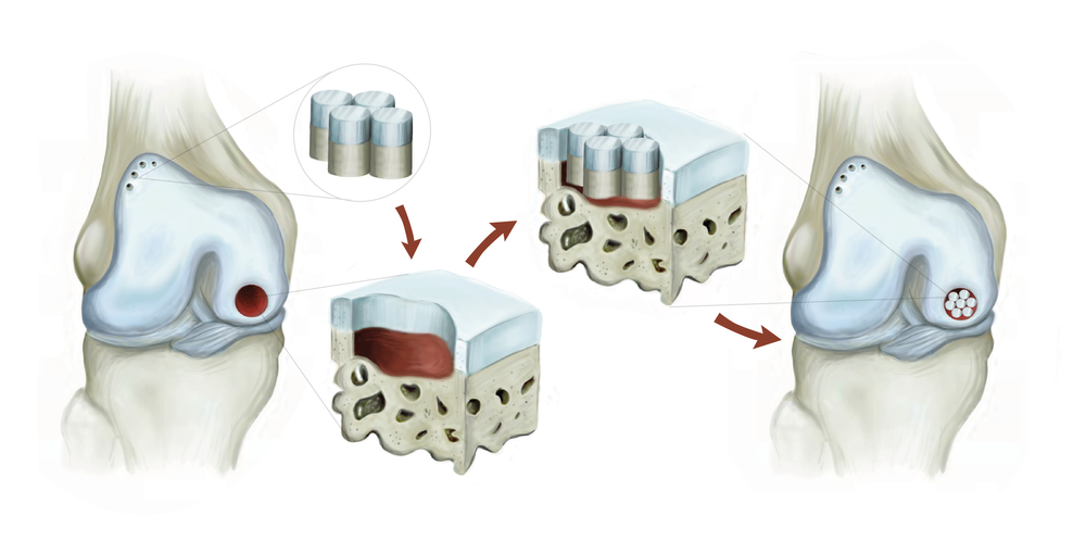
Osteochondral grafting of articular cartilage is a procedure of treating a damaged cartilage where the hidden bone is exposed. The method is usually executed for weight-bearing bones, including the knees. Even so, it might also be executed for other joints.

Covering the end bones of our joints all over our body, the articular cartilage is that white, smooth tissue.  What makes it easier and simple to move are the healthy cartilage in the joints.  It enables the bones to glide smoothly to each other with a bit of friction. Also, articular cartilage can be harmed by chronic diseases, like arthritis or injury.  

A full-thickness or limited cartilage erosion or injury of the cartilage caused by serious arthritis can lead to the vulnerability of the underlying bone. Hence, in cases of an injury in the cartilage, this surgery is done with the aim of achieving a painless range of movement and restoring knee strength.

TYPES

The two common types of osteochondral grafts are:

  • Allograft– This type utilizes a bone and cartilage from another area in the knee from the person’s body.
  • Autograft– A tissue graft is acquired from a knee donor and created to suit the patient’s knee.

When Should Osteochondral Grafts Not Be Performed?

The procedure might not be recommended for the following conditions:

  • Poor nutritional status
  • Severe underlying health conditions
  • Joint diseases
  • Tumors
  • Global arthrosis
  • Old people with chronic joint diseases, like osteoarthritis

TREATMENT

How Are Osteochondral Grafts Done?

Before the surgery, the doctor will perform a physical exam, blood test, and imaging tests to evaluate your joint condition. Furthermore, fitness for the procedure and consent is also acquired. 

Since there is a broad range of osteochondral disorders, the surgeon might do an arthroscopic evaluation to analyze chondral lesions prior to executing an osteochondral grafting. 

After The Procedure 

The patient can go out 2 to 3 days after the procedure is done. Weight-bearing is permitted, while an immobilizer is still worn for weight-bearing joints. 

The immobilizer may be taken off for a range of movement exercises and stationary activity during the first three weeks after the surgery. Furthermore, total recovery might take seven to eight weeks. 

Regular check-ups with the doctor and physical therapy would be needed. The doctor may also execute an arthroscopic evaluation to observe the healing of the joint.

Related Articles

TETRALOGY OF FALLOT

Overview and FactsTypes and SymptomsDiagnosis & MedicationsOverview and Facts Tetralogy of Fallot is a congenital heart defect that affects the [...]

TRICHINOSIS

Overview and FactsTypes and SymptomsDiagnosis & MedicationsOverview and Facts Trichinosis, also known as trichinellosis, is a parasitic infection caused by [...]

TRIGEMINAL NEURALGIA

Overview and FactsTypes and SymptomsDiagnosis & MedicationsOverview and Facts Trigeminal neuralgia is a neurological condition characterized by severe facial pain. [...]