MAMMOGRAMS
A mammogram is an X-ray imaging of your breasts designed to detect tumors and other abnormalities. It plays a key role in early breast cancer detection and help decrease breast cancer deaths.
A mammogram can be used either for screening or for diagnostic purposes. How often you should have a mammogram depends on your age and your risk of breast cancer. Risk factors include family history of breast cancer or a history of precancerous breast lesions. Talk with your doctor about your risk factors, your preferences, and the benefits and risks of screening. Together, you can decide what screening mammography schedule is best for you.
Risks
Risks and limitations of mammograms include:
- Mammograms expose you to low-dose radiation.
- Mammograms aren’t always accurate.
- Mammograms in younger women can be difficult to interpret.
- Having a mammogram may lead to additional testing.
- Screening mammography can’t detect all cancers.
- Not all of the tumors found by mammography can be cured.
How you prepare
To prepare for your mammogram:
- Choose a mammogram facility certified by the Food and Drug Administration. This will ensure that the facility meets certain standards.
- Schedule the test on the week before and the week during your period when your breasts are most likely to be tender.
- Bring your prior mammogram images.
- Avoid using deodorants, antiperspirants, powders, lotions, creams or perfumes under your arms or on your breasts. Metallic particles in powders and deodorants could be visible on your mammogram and cause confusion.
- To ease the discomfort of the test, you may take over-the-counter pain medication, such as aspirin, acetaminophen (Tylenol, others) or ibuprofen (Advil, Motrin IB, others), about an hour before.
During the test
- Wear a two-piece outfit that day so you can easily remove your clothing from the waist up. Also, remove your neck jewelry.
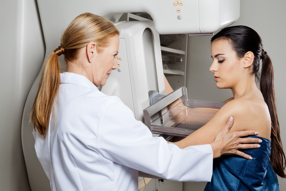
- For the procedure itself, stand in front of the X-ray machine designed for mammography. The technician places one of your breasts on a platform and raises or lowers the platform to match your height.
- Gradually, your breast is pressed against the platform by a clear plastic plate. This is for the X-ray to penetrate the breast tissue. Pressure is applied for a few seconds to spread out the breast tissue. The pressure may be uncomfortable or even painful but rest assured it isn’t harmful.
After the test/Results
- Wait while the technician checks the quality of the images.
- If you need not to repeat the test, you may dress and resume normal activity.
- A radiologist interprets the images and sends a written report of the findings to your doctor.
Possible findings include:
- Calcium deposits (calcifications) in ducts and other tissues
- Masses or lumps
- Asymmetric areas on the mammogram
- Dense areas appearing in only one breast or one specific area on the mammogram
- New dense area that has appeared since your last mammogram


