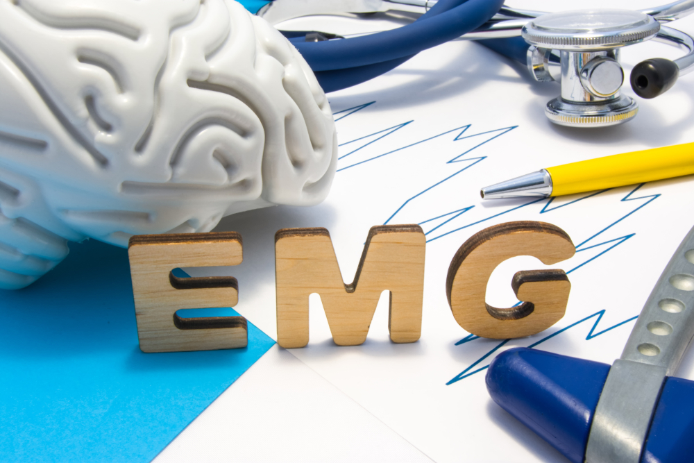
DIAGNOSIS
The process starts with a needle inserted into the skin to the muscle. The electrical activity identified by this needle serves as an electrode. These activities are shown visually on an oscilloscope and may also be identified audibly with a speaker.
Several needles are inserted at different locations to get an informative electromyogram (EMG). It is necessary because skeletal muscles are large. When the identified parts are inserted by needles, the patient will contract the muscle.
The existence, size, and shape of the waveform of the action produced on the oscilloscope give information on the ability of the muscle to respond to nervous stimulation. The contraction of muscle fiber produces an action potential. The growth of muscle fiber affects the rate of how frequently an action potential occurs and its extent.
What other test is conducted during an intramuscular electromyogram (EMG)?
A nerve conduction velocity (NCV) test is simultaneously conducted with an electromyogram (EMG). In NCV or nerve conduction velocity test, the nerve is electrically revived. The second electrode determines the electrical stimulation ‘down-stream’ from the first. It is done mainly with surface patch electrodes similar to those used on electrocardiograms that were placed on the skin on the nerve at different places. The electrode stimulates the nerve with a very gentle electrical impulse. The electrical activities are also recorded by the electrodes. To calculate the speed of impulse transmission or nerve conduction velocity; the distance between electrodes is determined, and the time it takes for electrical impulses to travel between electrodes is also obtained. Now, if the result has a decrease in the speed of transmission, it signifies a nerve disease.
The NCV test can detect nerve disorders such as neuropathy or conditions in which muscles are affected due to nerve injury, such as carpal tunnel syndrome. It is always imperative to maintain normal body temperature before the NCV test is conducted.
The intramuscular electromyography (EMG) method has a high distinction to an individual motor unit action potential. Dissection of intramuscular signals into individual motor unit action potentials consists of recognizing and classification, usually accompanied by separation of superimposed action potentials. An intramuscular electromyogram (EMG) signal is the tool used to examine physiological motor unit properties.
However, its utilization in clinical procedures is rare. It needs a human interaction and the problem in translating the quantitative data provided by electromyogram (EMG) signal decomposition serves as a basis in making clinical decisions. The most recent clinical utilization of intramuscular electromyogram (EMG) signal is on the diagnosis of myopathies- a disease caused by the alpha motor neuron. The neuromuscular link through the examination of the interference signal or the form of some motor unit action potentials, generally without a full decomposition of the signals.